Anatomical Name Of Lower Back Muscles / Lower Back Muscle Anatomy And Low Back Pain. Muscles of the lumbar spine. It is these ones that can be strained and fatigued and lead to your pain and spasms. The muscles of the lower back, including the erector spinae and quadratus lumborum muscles, contract to extend and laterally bend the vertebral column. The back muscles can be three types. Let us introduce you to each of these muscles presented in our diagram.
The extensors, which include the many muscles that attach to the spine and work together to hold your back straight while enabling you to extend it. Other extrinsic muscles are the: Your lats are a major back muscle and mover of your shoulder joint. Muscle anatomy study guide 12 photos of the muscle anatomy study guide anatomy and physiology muscle study guide, anatomy physiology muscle study guide, cat muscle anatomy study guide, muscle anatomy study guide, muscle study guide for anatomy, human muscles, anatomy and physiology muscle study. This muscle is the largest flexor of the foot.
Anatomy Of The Back Spine And Back Muscles Kenhub from thumbor.kenhub.com 1 your spine in this region has a natural inward curve. To perform clinical clinical orthopedic manual therapy to the lumbar spine. The back muscles can be three types. The muscles that move the upper legs (thigh) there are many muscles that move the large bone of the thigh. The flexors, which attach at your lumbar spine (lower back), and enable you to bend forward. The lumbar spine is the lower back that begins below the last thoracic vertebra (t12) and ends at the top of the sacral spine, or sacrum (s1). Muscle names of lower back. The flexor muscles are attached to the front of the spine and enable flexing, bending forward, lifting, and arching the lower back.
Intrinsic muscles (figure 5.2) are the small and firm muscles that travel along each of your vertebra from the pelvis to the cranium.
Its name means belly of the leg,and its common name is the calf muscle. See back muscle anatomy stock video clips. The quadratus lumborum muscles (orange, in the image above) are found in the lower back (also called the lumbar area). 1 your spine in this region has a natural inward curve. Related posts of muscles of the lower back and buttocks diagram muscle anatomy study guide. The muscles of the back can be arranged into 3 categories based on their location: It is innervated by anterior rami of spinal nerves, reflecting its embryological origin outside the back. There are three different muscle groups found in the back: Muscles of ulnar nerv 12 photos of the muscles of ulnar nerv muscles of ulnar nerve, muscles the ulnar nerve stimulates, muscular branches of ulnar nerve, human muscles, muscles of ulnar nerve, muscles the ulnar nerve stimulates, muscular branches of ulnar nerve Deep back muscles superficial back muscles action movements of the shoulder. Superficial back muscles, intermediate back muscles and intrinsic back muscles.the intrinsic muscles are named as such because their embryological development begins in the back, oppose to the superficial and intermediate back muscles which develop elsewhere and are therefore classed as extrinsic muscles. Human musculature bodybuilding infographic muscular system vector human anatomy back muscle anatomy bicep male muscular anatomy human body anatomy female female anatomy muscle hamstrings muscle. It is composed of trapezius, latissimus dorsi, rhomboid major, rhomboid minor and levator scapulae.
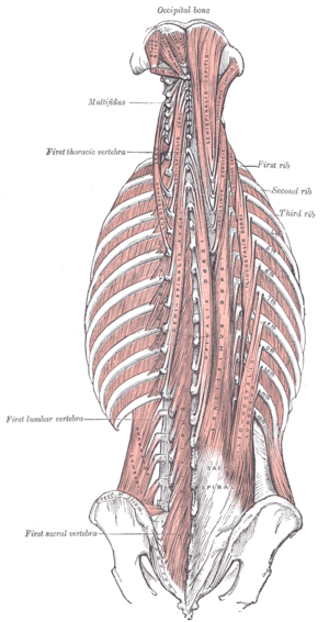
Muscles of the back can be divided into superficial, intermediate, and deep group. The vertebral column of the lower back includes the five lumbar vertebrae, the sacrum, and the coccyx. This curve, called lordosis, helps to: Related posts of muscle names of lower back muscles of ulnar nerv. 7 / 10 (1 vote) muscles of lower back diagram in this image, you will find an occipital bone, sternocleidomastoid, trapezius, deltoid in muscles of the lower back diagram.
Lower Back Muscle Anatomy And Low Back Pain from ix-cdn.b2e5.com The extensors, which include the many muscles that attach to the spine and work together to hold your back straight while enabling you to extend it. The vertebral column of the lower back includes the five lumbar vertebrae, the sacrum, and the coccyx. Its name means belly of the leg,and its common name is the calf muscle. The muscles of the back that work together to support the spine, help keep the body upright and allow twist and bend in many directions. Lay on your back with your arms at either side. 7 / 10 (1 vote) muscles of lower back diagram in this image, you will find an occipital bone, sternocleidomastoid, trapezius, deltoid in muscles of the lower back diagram. Intrinsic muscles (figure 5.2) are the small and firm muscles that travel along each of your vertebra from the pelvis to the cranium. The muscles of the back can be arranged into 3 categories based on their location:
The quick answer to this question is the muscles of the lower back are the multifidus, longissimus, spinalis, and quadratus lumborum.
Located at the front of your body, the flexors. Deep back muscles superficial back muscles action movements of the shoulder. The back consists of the spine, spinal cord, muscles, ligaments, and nerves. On this page, you'll learn about each of these muscles, their locations and functional anatomy. To perform clinical clinical orthopedic manual therapy to the lumbar spine. Muscles of the back can be divided into superficial, intermediate, and deep group. Intermediate extrinsic muscles of the back: The back muscles can be three types. The extensors, which include the many muscles that attach to the spine and work together to hold your back straight while enabling you to extend it. Its name means belly of the leg,and its common name is the calf muscle. Lumbar spine lower back and superficial muscles the muscles of the lower back help stabilize, rotate, flex, and extend the spinal column, which is a bony tower of 24 vertebrae that gives the body. Intermediate back muscles and c. These muscles include the large paired muscles in the lower back, called erector spinae, which help hold up the spine, and gluteal muscles.
Other extrinsic muscles are the: See back muscle anatomy stock video clips. Intrinsic muscles (figure 5.2) are the small and firm muscles that travel along each of your vertebra from the pelvis to the cranium. The superficial group, also known as the appendicular group, is primarily associated with movement of the appendicular skeleton. (2017, elsevier) should be consulted.
Transversospinales Physiopedia from www.physio-pedia.com The quadratus lumborum muscles (orange, in the image above) are found in the lower back (also called the lumbar area). There are three different muscle groups found in the back: Your lats are a major back muscle and mover of your shoulder joint. These muscles provide posture and stability to the body by holding the vertebral column erect and adjusting the position of the body to maintain balance. This muscle is the largest flexor of the foot. Muscle names of lower back. The pelvic floor muscles also help increase this pressure, which provides stability to the spine and trunk. Most of the time, back muscle pain is diagnosed then treated with little more than a prescription of rest, painkillers and muscle relaxants.
The back consists of the spine, spinal cord, muscles, ligaments, and nerves.
See back muscle anatomy stock video clips. The superficial back muscles include the suboccipital muscles, trapezius, latissimus dorsi, levator scapulae, rhomboids and serratus posterior muscles. The back anatomy includes the latissimus dorsi, trapezius, erector spinae, rhomboid, and the teres major. Most of the time, back muscle pain is diagnosed then treated with little more than a prescription of rest, painkillers and muscle relaxants. The muscles of the back can be arranged into 3 categories based on their location: Serratus posterior superior and serratus posterior inferior muscles. Intrinsic muscles (figure 5.2) are the small and firm muscles that travel along each of your vertebra from the pelvis to the cranium. The back muscles can be three types. Tutorial and quizzes on skeletal muscular anatomy. The quick answer to this question is the muscles of the lower back are the multifidus, longissimus, spinalis, and quadratus lumborum. Muscle names of lower back. These structures work together to support the body, enable a range of movements, and send messages from the brain to. Serratus posterior inferior and superior.